
Wrinkles and folds on the brain
 One of the first things people notice about the human brain is how convoluted its surface is. Instead of being smooth and nearly featureless like a kidney or spleen, the cerebral cortex (the thin layer of gray matter forming the outer surface of the brain) is chock-full of wrinkles and folds. Click on the image above for a larger view.
Technically, each crevice is called a sulcus (pl. sulci) and each ridge between the crevices is called a gyrus (pl. gyri). Sulci and gyri are simply a way of increasing the surface area of the cerebral cortex (and therefore the number of neurons) without greatly expanding the size of the skull. Good thing, too - if our skulls were much bigger, they wouldn't be able to squeeze through the birth canal and C-sections would become the norm. Not to mention that those enormous heads would make us look like Hollywood aliens.
There are perhaps 30 or so named sulci and gyri, but learning to identify them - a time-honored task of every medical student - isn't as easy as it might sound. It isn't true that if you've seen one brain, you've seen them all. Although the total surface area of the cortex is roughly the same in all people, there are large variations in the size of particular areas. Whether these differences in area are related to differences among individuals in various skills and functional capacities is largely unknown (but there have been a few intriguing studies, such as this one and this one).
For the sake of all those sleep-deprived med students, why can't we have a brain that looks more like, say, a howler monkey's?
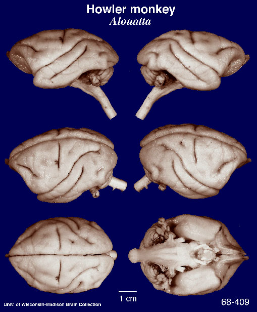 So nice and smooth, almost no sulci or gyri to memorize. Actually, we humans start out wrinkle-free early in development. Check out this brain from a fetus at 22 weeks:
 As neurons continue to divide, grow, and migrate, the cortex folds in on itself, forming a recognizable but unique pattern of bumps and grooves. Unless you happen to have lissencephaly. Children born with lissencephaly (which means "smooth brain") are severely retarded and many die before the age of 2.
So we should be happy with our brain wrinkles. As I've often pointed out to my students, it could be worse. We could be dolphins:

Finally, here's a quiz. Compare the brain below to the brain at the top of this post. What's wrong with it?
 Answer: nothing, if you're a chimpanzee. Clearly, there are anatomical differences between a chimpanzee brain and a human brain, particularly in the size of the prefrontal cortex (the front part of the cerebral hemispheres). But the similarities in brain anatomy, like the similarities in DNA, are striking.
All but one of the brains shown here belong to the University of Wisconsin and Michigan State Comparative Mammalian Brain Collections. I modified the first and last images slightly to make them easier to compare. The fetal brain came from humanpath.com.
posted by Brad @ 5:40 AM
28 comments
![]()
![]()


28 Comments:
Thanks Brad- I wanted to know if chimpanzee have
prefrontal lobes?
I'm not a med student!
Yes, chimpanzees and other primates have a prefrontal cortex.
I learned recently that whether humans have a disproportionately large prefrontal cortex is still an open question. Surprisingly, one recent MRI study concludes that the human frontal cortex is not larger than expected for a primate brain of human size. The authors suggest that "the special cognitive abilities attributed to a frontal advantage may be due to differences in individual cortical areas and to a richer interconnectivity, none of which required an increase in the overall relative size of the frontal lobe during hominid evolution."
However, they also acknowledge that their study doesn't settle the question about the prefrontal cortex, since they measured the entire frontal cortex, which includes the prefrontal cortex and a few other areas. The problem is that the prefrontal cortex has no clear anatomical boundaries; you have to look for differences at the microscopic level. Given the formidable challenges (scarcity of great ape brain material and cost), no one has attempted such a study for the entire prefrontal cortex. One cytoarchitectonic study shows that Brodmann area 10 is relatively large in humans compared to apes, but area 10 is only part of the prefrontal cortex.
Which parts did you modify?
Good question - I just moved the chimpanzee brains closer to each other so that the aspect ratio of the chimpanzee image would be nearly the same as the aspect ratio of the human image.
But why are they wrinkled? I can see that they are, and that it is to increase surface area, but what is the benefit of that? Couldn't you pack more neurons in a sphere?
Not sure, sara. The brain is organized so that most of the gray matter lies superficially, while white matter lies at deeper levels. One idea is that wrinkling is the most straightforward way to keep the gray matter near the surface without completely rewriting the developmental program.
So is there any functional advantage to keeping the gray matter near the surface? Since gray matter consumes a disproportionate amount of energy compared to white matter, it might make sense to keep gray matter closer to the major arteries of the brain, which all course along the surface.
How deep are the folds of the brain?
Hmm...I'd say half an inch is a typical depth, but they range from very shallow (1/8 inch) to deep (one inch or more).
Omggg (oh my gosh goly goodness) I'm reading this to my cuzan who is nine years old,and i want to know what her brain would look like but I'm out of luck. it want even easy for her to understand, which is understandable, i barely knew myself. I'm twelve and think this stuff is interesting, just confusing, same with her.
Hey Brad, I am actually working on the issue with the size of prefrontal cortex in the chimpanzee. I have measured the total and prefrontal surface areas in a sample of chimpanzee brains. So far I have been having trouble finding published values for the total surface area and the prefrontal surface area in the human brain. Would you happen to know of a source for these values?
Great question, knight816. A 1996 MRI study (http://www.pubmedcentral.nih.gov/articlerender.fcgi?artid=1167579) came up with an estimate of about 1600 cm2 for the total surface area of the brain. Finding an estimate for the prefrontal cortex will be harder - see the second comment above for more info. This weekend I'll try to track down the papers I mentioned in that comment, since the links to them appear to be broken now.
Below are references for the articles I mentioned above. You may be able to find the complete articles (pdf) online if you google the titles.
Semendeferi K, Lu A, Schenker N, Damasio H. Humans and great apes share a large frontal cortex. Nat Neurosci. 2002 Mar;5(3):272-6. (http://www.nature.com/neuro/journal/v5/n3/abs/nn814.html)
Semendeferi K, Armstrong E, Schleicher A, Zilles K, Van Hoesen GW. Prefrontal cortex in humans and apes: a comparative study of area 10. Am J Phys Anthropol. 2001 Mar;114(3):224-41. (http://www3.interscience.wiley.com/journal/77004262/abstract)
Hope that helps!
Brad
So the oonly reason we have crevices in our head is so it can cover more area in our skull.
What is the surface area of the pia mater vs. choroid plexus?
I don't know. I'd guess the surface area of pia mater is at least 3 or 4 times that of choroid plexus, maybe more...
''Wrinkles and folds on the brain'' - sounds like we have a bit of origami of the brain.
;-)
glad you popped in on theme thursday...really fascinating stuff...
Hey! I was just commenting on Paul C's blog about wrinkles on our brains and came here next! De Ja Vue!!! Glad you joined TT today...come back! :)
scientific stuff,
amazing knowledge!
To Chad,
do you know if it is true that everytime you learn something new your brain grows a wrinkle.
from Jessie from ODC.
Im not a medical student but i am taking an Anatomy/Physiology class in college..I don't understand the difference between a gyrus, sulcus and a fissure...if you could please explain that would be so helpful! Thank you!
A fissure is an especially deep or prominent sulcus. Sulcus and gyrus are defined above in the 2nd paragraph of the essay. Hope that helps!
Thank you!!
Not sure what you meant by at least we don't have dolphin brain. What are you are saying about dolphins brains?
Dolphin brains are really wrinkly.
Is there any difference in the function of the gyri and sulci
I'm curious For the answer to Jessie Parkers question (kind of why I'm here) ^-^ I'm into psychology and the brain.
hi! can you please tell me whether the cerebrum or the cerebellum has more number of folds and why?
Post a Comment
<< Home